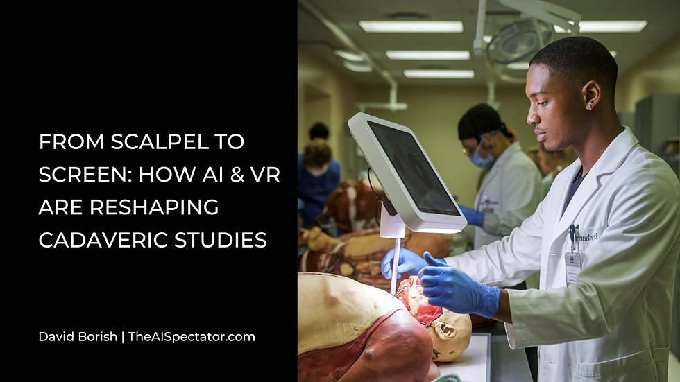
A recent study published in Cureus on October 16, 2024, titled "From Scalpel to Simulation: Reviewing the Future of Cadaveric Dissection in the Upcoming Era of Virtual and Augmented Reality and Artificial Intelligence," has ignited fresh discussions about the future of anatomical education. This timely review explores the intersection of traditional cadaveric dissection (CD) and emerging artificial intelligence (AI) technologies in medical training.
Cadaveric dissection, a practice dating back to the 3rd century B.C., has long been the gold standard for teaching human anatomy. It provides medical students with irreplaceable hands-on experience, offering insights into the intricacies of the human body that textbooks alone cannot convey. The tactile and spatial understanding gained through CD has been crucial in developing the skills necessary for clinical practice and surgery.
However, the landscape of medical education is rapidly changing. AI-driven tools, virtual reality (VR), and augmented reality (AR) are making significant inroads into anatomy curricula. These technologies offer highly detailed 3D models, interactive simulations, and the ability to explore anatomical variations that might be rare in cadaveric specimens.
Platforms like Anatomage and Microsoft's HoloLens are providing immersive experiences where students can virtually dissect and study anatomy with unprecedented flexibility.
The Cureus study highlights the potential benefits of integrating these AI technologies into anatomy education. These include increased accessibility, personalized learning experiences, and improved knowledge retention. For instance, AI algorithms can generate detailed, manipulable 3D models of the human body, allowing students to explore anatomical structures in ways previously impossible.
Yet, as the study points out, AI and VR are not without limitations. They lack the tactile feedback crucial for developing surgical skills and understanding tissue textures. The unpredictability and complexity of real human anatomy can't be fully replicated by even the most advanced AI models. Additionally, the ethical dimension of working with human remains, which many argue instills a sense of professionalism and respect in future doctors, is absent in digital simulations.
The research suggests that the future likely lies in a hybrid approach. By integrating AI with traditional dissection, medical educators could enhance pre-dissection preparation, offer real-time guidance during hands-on sessions, and provide supplementary learning experiences. This combination aims to create a more comprehensive educational experience, leveraging the strengths of both methods.
The study also raises important questions about the role of CD in developing surgical skills. While AI can simulate complex procedures and offer risk-free practice environments, it currently cannot replace the experience of working with real human tissues. The muscle memory and confidence required for successful surgical outcomes are still best developed through hands-on practice.
As we move forward, the Cureus review emphasizes the need for ongoing research to evaluate the effectiveness of these hybrid models. The goal is to ensure future healthcare professionals receive a well-rounded education that combines the irreplaceable insights gained from cadaveric dissection with the innovative capabilities of AI technology.
While AI is transforming medical education, the study suggests that cadaveric dissection is here to stay. The challenge lies in finding the right balance to produce skilled, knowledgeable physicians ready for the complexities of modern healthcare. As medical institutions navigate this evolving landscape, they must consider how to best integrate these technologies while preserving the unique benefits of traditional anatomical education.
_edited.png)


Kommentare